“I have searched through the literature, and have found no other mention of a magnetic fibrous capsule (or indeed any magnetic biological tissues) whatsoever.” ~ Dr. Muirhead
Alien Implant Mystery – Part 2
The opportunity to study and document for a TV series what we suspected may be a non-terrestrial implant based on the conscious personal account of Natalie along with what we learned from her hypnotically induced session was a fascinating process, shared in the previous post; Alien Implant Mystery.
Since that report I have learned a few more details and have discovered a new anomaly that remains to be explained scientifically.
Continuing with what was learned after use of Haematoxylin and Eosin to stain tissue sections of the object removed from Natalie’s upper right arm, photomicrographs and graphics were provided by the university laboratory that analyzed tissue samples taken from Natalie’s arm. What was found in that sample is a very active fibrotic capsule.
The question then arises, which has yet to be explained by the scientists involved thus far:
Why are new blood vessels, encapsulated within a 25-year-old hard skin fragment, highly active and why did it attract neodymium magnets prior to the surgical removal of that skin fragment?
What I learned while shooting the scientific analysis is presented here, by the pathologist on camera:

“The stain combination used was Haematoxylin and Eosin. It is by far the most common tissue stain for preliminary pathological examination. If we’d known about the bone ahead of time, we probably would have used something that stains mineralized bone and osteoid seams, osteoblasts, and osteoclasts (bone cells). I won’t get into the specifics, but haematoxylin generally binds to the nuclei of cells and stains them blue, while eosin generally stains protein rich structures some shade of pink.
The dark spots are, in almost every case, the nucleus of a cell. You can tell what type of cell based on the morphology, size, and number of nuclei. Most of the nuclei in your image are probably fibroblasts. The ones that appear very dark are probably where the tissue has folded onto itself during sectioning, and there is more than one layer present. More importantly, however, is that there doesn’t seem to be any immune cells (white blood cells) present anywhere. They are typically what you look for in these types of cases, as they would tell you if the body is fighting this foreign material, or, like in your case, ignoring it. Attached is a picture of a very active fibrotic capsule from one of my experiments (day 20). The oval object is a collagen-based biomaterial I have injected under the skin of a mouse. You can see, from a distance, that there is a purple/blue ring around it. That area is so active will infiltrating cells, it looks blue. It is also absolutely packed with new blood vessels. It is also almost half a millimeter thick, as opposed to your capsule, which is more like 20 microns thick.
The purplish-red tissue is what was stained with eosin, which primarily stains amino acids (proteins). The pink blob in the middle labeled D is a chunk of cartilage. It’s a little blurry because it’s not flat against the slide – it has some height, which prevents proper focus. The pinkish stuff labeled A is skin. It was probably removed attached to the object during the surgery. It’s dermal tissue. The rest of the purple stuff is the collagen capsule that surrounded the object, and some bits of associated dermal tissue.
The greyish smudges are on the slide. They are bubbles from the sample not having enough time to set properly, and appear above the plane of focus.
The foreign material was determined to likely have been bone. I do not want to appear too categorical without more thorough testing.
There are very few capillaries in the image. I have circled one of them and labeled it C. If you look closely at C, you can see a ring of darkly staining endothelial cells, and red blood cells (erythrocytes) inside. There are a few other examples, but not many. It’s hard to say how unusual that is. Pathology on a fibrotic capsule this old is extremely rare. I have nothing to compare your sample with.
In my work, I see blood vessel formation all the time. However, when a material is tolerated by the body (as yours was ie no immune cells), and has been encapsulated for a sufficient period of time (say 3 months), most of these blood vessels are resorbed and disappear. I was speaking intuitively when I said I was not expecting to find any. If most are gone after 3 months, surely there ought to be none after 25 years! I have no better answer to give. If you want a quote, I will commit to my previous statements that blood vessels were not expected, and I was surprised to find them.
The body is an extremely complicated landscape of competing signals. It’s impossible to point to one microscopic feature and describe its lineage with any clarity. Just as a point of clarification – the blood vessels are inside the capsule, and do not directly interact with the bone fragment within.
“Osteogenic in nature” refers to the material inside the capsule.
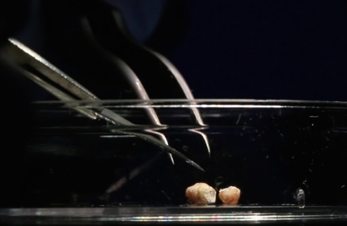
Without callipers, I’d say we cut about half way through. The object was around 10mm long. We split it into two halves: one that was 6.5mm and one that was 3.5mm. The image you have is from the larger piece, and I think is likely from ~3mm-3.5mm inside.
I seriously doubt attempting to section deeper would discover anything new – but I suppose you never know.
While handling the object, and later cutting through it, I noticed it was hard. Because it was too small to see, metal was a reasonable assumption at the time. If it was a soft metal, like a lead or pewter bb-pellet, or some pencil lead, it may have cut in a similar way. Lead and bone both have a hardness of ~2 on the Mohs scale. Bone fits my observations as well – it just hadn’t occurred to me until the SEM results came it.
Bone requires specific preparation to section. It needs to be decalcified with a strong acid (usually hydrochloric acid) solution for several days. Once complete, bone will become almost rubbery, and can be sectioned successfully. This would not have been possible with the time constraints in place, and we did not know there was any bone until after testing. The sections we got turned out remarkably well all things considered.” ~ Ben Muirhead, PhD, Department of Biomedical Engineering

Q&A
Sid: “The event of awaking in the middle of the night with a shooting pain at the age of 8 or 9 years old to suddenly feel this tiny bump at the skin’s surface. How can it suddenly appear?”
Dr. M: Perhaps a previously dislodged bone fragment pierced the skin from underneath, causing the acute pain sensation she describes. From there, the bone fragment may have been encapsulated and remained more or less quiescent until it was removed.
Sid: “How can this be normal bone or cartilage as both do not attract magnets?”
Dr. BM: Unknown. We can only report what was discovered.
Sid: “How could a piece of bone remain just below the skin’s surface and remain in the same place for 20-plus years length of time?”
Dr. BM: Once a foreign body has been encapsulated, it can remain that way ad infinitum. This is a link to a paper describing the foreign body response in some detail. It is a huge problem for implanted medical devices, because the body can wall them off and cause device failure even if they are meant to help (ie an insulin pump or prosthetic hip). Devices that have been rendered inoperable in this way often remain in the patient for the remainder of their life, as the effort to remove them is not worth the trauma of surgery, and they are completely inert while encapsulated. Your bone fragment may be no different.
Sid: “Why was no bone or tissue growth that occurred in over 20 years, especially if there was blood vessels feeding the area?”
Dr. BM: Materials encapsulated in collagen and locked within the skin often remain there, as yours did, for life, with no deleterious consequences. As I will mention in my report, your sample did contain an unexpectedly active capsule. Usually, the foreign body response is resolved one way or another after about 12 weeks. Either everything returns more or less to normal, or a chronic, painful immune reaction will persist until something is done to treat it. That being said, this type of pathological result exists on a spectrum, where ‘normal’ is simply the most common case. These results are still well within a ‘normal’ range of expected tissue responses.
Sid: “There was no bone or tissue growth that occurred in over 20 years. Is this something we would have expected?”
Dr. BM: If this was a fragment chipped from a nearby bone, it would not grow. If it was something more exotic, like some kind of benign fibroma or hard tissue sarcoma, it is impossible to predict how they will grow or not grow.
Sid: “How is the response to magnetism explained and the reaction of heat when a piece of copper was placed over the object. Why would bone have these reactions?”
Dr. BM: Again, bone would not react in these ways. I have no plausible explanation for these phenomena.
Sid: Do you believe that is possible and if so, would you have seen any residual metal elements in your pathology analysis?
Dr. BM: This is as likely an explanation as any I can think of. I didn’t see any metal, so they answer is no in this case.
Sid: Would you normally expect to see active white blood cells and what role would they play in a foreign object?
Dr. BM: White blood cells indicate a persistent immune response. Generally speaking (as I’ve mentioned before), encapsulated objects like this one typically fall into one of two categories: either they react with the immune system and create inflammation (redness, itching, swelling, pain, etc) or they are walled off harmlessly. Yours seems to fall into the latter category, despite the odd blood vessel. If the host of this material was comfortable, I would not expect to see any immune cells – and we didn’t.
Sid: If the blood vessels do not link and interact with the bone fragment within, where are they connected to and what are they interacting with? Do they actively link and interact to the rest of the body outside the capsule? If they are not directly interacting with the bone, is the bone live or dead, or something else?
Dr. BM: The blood vessel was interacting with the capsule. During its formation, many new blood vessels form to bring raw material to the site of the object to wall it off. They are likely connected to the cardiovascular system.
Dr. BM: I did not get a clean enough section of the interior to say if the bone/cartilage within was ‘alive’ or ‘dead’. My guess would be alive, although bone is mostly made of a ‘dead’ mineral matrix, and contains very few cells outside of the marrow.
Dr. BM: I have searched through the literature, and have found no other mention of a magnetic fibrous capsule (or indeed any magnetic biological tissues) whatsoever. I’m sorry I don’t have a better answer for you! – BEN MUIRHEAD, PhD, Department of Biomedical Engineering

Additionally, Dr. Michael D. Noseworthy, Ph.D., P.Eng. Co-Director, McMaster School of Biomedical Engineering, Associate Professor, Department of Electrical and Computer Engineering, McMaster University oversaw XRF analysis.
X-ray Fluorescence (XRF) and X-ray Diffraction (XRD) Analyzers provide qualitative and quantitative material characterization for detection, identification, analysis, quality control, process control, regulatory compliance, and screening, for metals and alloys, mining and geology, scrap and recycling, environmental and consumer safety, education and research, and general manufacturing.
The test revealed that the sample had copper at about 23 ppm. Nickel was 95ppm. No platinum or gold. There was no silver in the arm sample. Below is a screen shot of the full results.

High Strangeness Conclusions:
- What we learned from this analysis is that the anomaly of why a small neodymium magnet attached to the arm of Natalie remains a mystery.
- Why there was an active blood flow to the encapsulated detached object beneath Natalie’s skin also remains inconclusive.
- What remains even more outstanding is what happened to this sample afterwards, to be presented in the next update of the Alien Implant Mystery – What is this teardrop shaped crystalline object?
If any EMN readers have scientific training and can offer insight as to what might explain why magnets were attracted to Natalie’s arm where the tissue was found, as well as why there was still an active blood flow to an isolated and encased chip of skin, please send in your comments below or write us at info@EarthMysteryNews.com.
…end of Part 2, Alien Implant Mystery – Anomalous Results Present
…PART 3 upcoming: Alien Implant Mystery – What is this teardrop shaped crystalline object?
Copyright 2014 images on videotape by Sid Goldberg, TV Director and EMN Publisher.
2 comments






















2 Comments
James
March 7, 2016, 3:39 amInteresting article. Have you compared the elemental readout of the test material with a baseline that most other people have?
REPLYSid Goldberg@James
March 7, 2016, 3:42 amJames, that would be an excellent test for us to do. Finding the right scientist to do so is what I need to research. In another article I will be writing, we have another sample that tested differently and the results are highly unusual, if not strange. In Part 3 I have images of something that has “appeared” on the test slides that is crystalline looking and highly strange. Look for that soon!
REPLY